





How to Fix Diastasis Recti

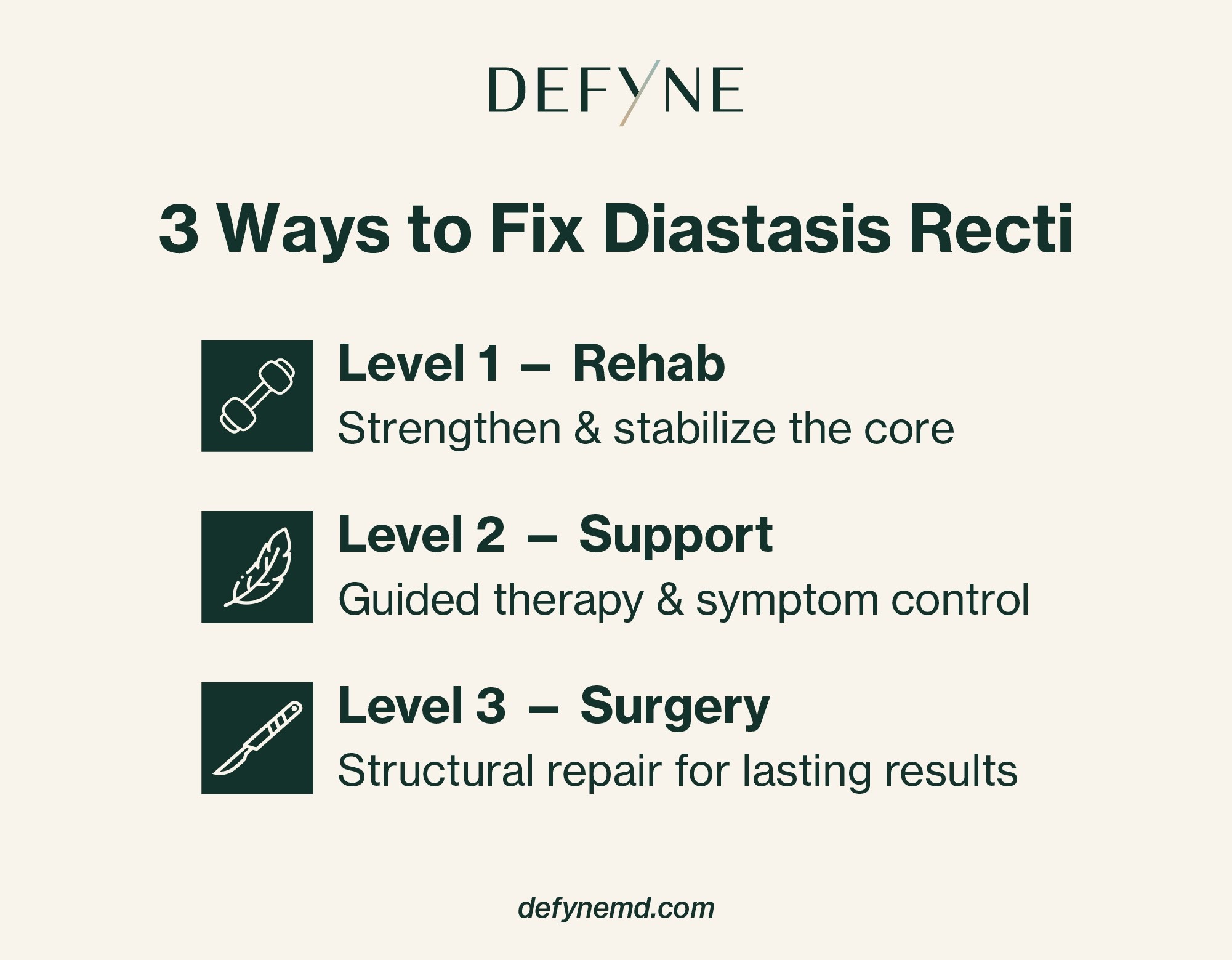
To fix diastasis recti, most people start with targeted core rehabilitation and, if bulging, weakness, or excess skin persists, move on to surgical repair. This can include plication—a minimally invasive technique—or a tummy tuck for diastasis recti.
Below, we walk through what diastasis recti is, why it happens, how it’s diagnosed, and how each treatment option stacks up.
Think you may have diastasis recti? Reach out to schedule a consultation to review your symptoms, exam findings, and options—from targeted physical therapy to minimally invasive repair to tummy tuck (abdominoplasty) when appropriate.
Non-Surgical Diastasis Recti Treatment
For some people, the best first step is a non-surgical rehabilitation plan focused on improving how the core functions. While exercises may reduce the separation in some mild cases, conservative treatment is usually aimed at decreasing doming and bulging, improving strength and stability for daily activities, and reducing symptoms.
- Pelvic floor therapy/physical therapy: Evaluation of core and pelvic floor coordination, plus an individualized home program to build strength.
- Breathing and pressure management: Training to reduce excessive abdominal pressure (often using exhale-based bracing) to minimize midline doming during movement.
- Support garments (optional): A binder or supportive garment may improve comfort early postpartum, but it doesn’t replace rehabilitation.
How to Fix Diastasis Recti With Surgery
Surgery can be a good option for patients who still have significant bulging, doming, core weakness, or day-to-day limitations after a structured rehabilitation program—and after pregnancy-related changes have stabilized.
The goal of surgery is to restore midline support by tightening the stretched connective tissue and bringing the rectus muscles back toward the midline—sometimes also addressing a coexisting hernia or excess skin, depending on your anatomy and goals.
- Open plication (functional repair): An incision-based repair that sutures (plicates) the linea alba/rectus sheath to narrow the gap and improve abdominal wall support.
- Abdominoplasty (“tummy tuck”) with plication: Combines midline tightening with removal of excess skin/soft-tissue laxity when contour concerns are also a priority.
- Minimally invasive repair: Smaller incisions for selected candidates who primarily want functional repair and do not need significant skin removal.
- Combined diastasis + hernia repair (± mesh): If an umbilical/ventral hernia is present, the plan may include hernia repair and, in some cases, mesh reinforcement based on tissue quality and defect size.
The “best” option depends on symptoms (function), anatomy (gap location, tissue quality, hernia), aesthetic goals (skin excess), recovery time, and scarring preferences.
The Defyne Approach to Diastasis Recti
When you’re weighing surgical options for diastasis recti, the choice usually comes down to how much skin laxity you have and how much downtime you can afford.
Defyne offers traditional tummy tuck surgery for diastasis recti, but treatment isn’t one-size-fits-all.
Tummy Tuck Surgery
A board-certified Defyne plastic surgeon will determine the best surgical technique for your anatomy and aesthetic goals. There are several types—mini, tunneled, traditional, reverse, and extended.
Traditional tummy tuck surgery addresses moderate to significant excess skin and can resolve diastasis.
No matter which approach you choose, we recognize that no two bodies or abdominal concerns are the same and work with you to ensure a safe, successful outcome.
What is Diastasis Recti?
In simple terms, diastasis recti is a separation in the middle of the abdomen between the left and right “six-pack” muscles, called rectus abdominis. When these muscles sit farther apart than usual, you might notice midline “doming” or a ridge when sitting up, accompanied by a persistent lower belly bulge.
This muscle weakness can also affect the feeling of core strength and stability, and sometimes lead to back discomfort.
Diastasis recti happens commonly during pregnancy, but also persists post-pregnancy. While this is a normal part of pregnancy, when it remains after pregnancy, it can be undesirable and affect appearance, quality of life, and self-esteem.
One study noted that as many as 33% of women experience diastasis recti at 12 months postpartum. It’s also common in women who have had multiple pregnancies, and it frequently remains present later in life.
Risk Factors for Diastasis Recti
During pregnancy, the growing uterus stretches the rectus abdominis muscles. Between these muscles lies a tough band of connective tissue called the linea alba. This tendon-like seam also stretches and thins, allowing the muscles to separate.
A few risk factors can make diastasis recti more likely, aside from pregnancy itself:
- Multiple pregnancies
- C-section
- High BMI
Once diastasis recti occurs, diastasis recti repair may be considered if symptoms persist—especially if you have ongoing bulging, weakness, or functional limitations despite a structured rehabilitation program.
How to Diagnose Diastasis Recti
If you think you may have diastasis recti, there are a few ways it can be diagnosed by a healthcare professional. You can start with an at-home screening to check for common signs of separation and decide whether it would be helpful to schedule a formal exam for a more accurate diagnosis.
At-Home Screening Steps
- Lie on your back with knees bent and feet flat.
- Place your fingertips across the middle of your stomach near the belly button.
- Lift your head and shoulders a few inches.
- Notice whether you see or feel a ridge or doming along the midline, and how many finger-widths fit between the left and right rectus muscles.
A gap of two finger widths or more (roughly 2 cm) is often used as a practical reference that diastasis recti may be present.
Clinical Diagnosis
A clinical diagnosis usually starts with a brief history and a hands-on abdominal wall exam.
Your clinician may ask when you first noticed a bulge, whether it changes with sitting up or lifting, and whether you have symptoms such as core weakness, back discomfort, pelvic floor symptoms, or a sense of “instability” during daily activities.
They’ll also review pregnancy and delivery history, prior abdominal surgery, and any history of hernia.
An important part of the visit is ruling out (or identifying) an umbilical or ventral hernia. Diastasis recti is a stretching and thinning of connective tissue, whereas a hernia involves a true defect in the abdominal wall that can allow tissue to protrude.
A clinician will check for a discrete opening, tenderness, or a bulge that behaves like a hernia (e.g., a lump that is not easily reducible).
A clinician will also estimate or measure the space between the rectus muscles, either by palpation or with tools such as ultrasound, which is considered the most accurate clinical tool for this measurement.
CT or MRI isn’t routinely needed for diastasis alone, but it may be used when a hernia is suspected, when symptoms are atypical, or for surgical planning.
Contact Defyne Today
Ready to discuss your options for diastasis recti treatment? Contact Defyne to schedule a consultation and receive a personalized plan tailored to your goals.
Contact Us
Request a Consultation
Are you ready to take the first step toward defining your destiny and achieving your best look yet? At Defyne, our board-certified plastic surgeons are skilled in performing both surgical and non-surgical procedures, achieving individualized, natural-looking results. Contact Defyne today to schedule your consultation appointment.